Study of a model for a rare genetic neurodevelopmental disorder revealed deficits of vasopressin in brain, which treatment restored neurophysiology and social behavior.
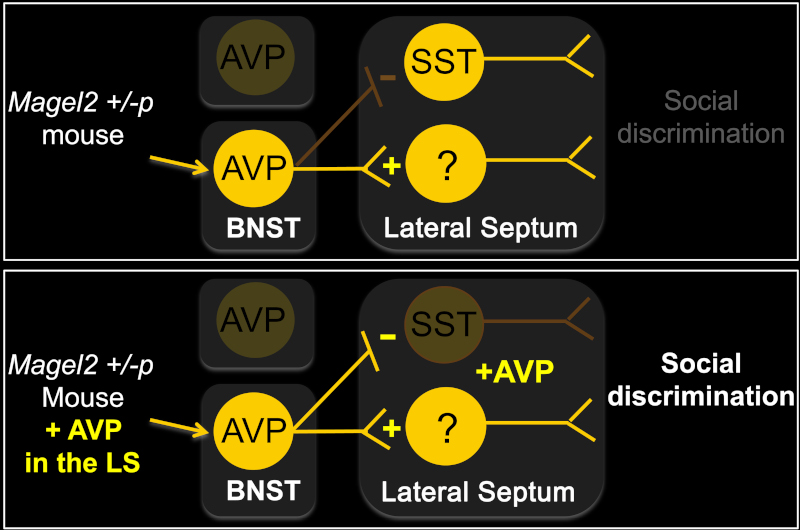
A collaborative study between IGF (Jeanneteau’s team) and teams at INMED and in the USA show an improvement of social engagement by a treatment with vasopressin or by the optogenetic activation of vasopressin neurons projecting in the lateral septum of autism mouse model. These treatments promoted the inhibition of somatostatin inhibitory neurons as well as the activation of other GABA neurons yet to be characterized.
Anatomic and functional disorganization of vasopressin neurons in individuals carrying a loss of function mutation in the MAGEL2 gene caused a deficit of neuropeptide secretion in the lateral septum latéral when somatostatin neurons are activated. First of all, this mechanism fail to activate when encountering unknown conspecifics. Then, the vasopressin neurons engaged in such response of indivuals carrying the mutation were unexpected. It is the extended amygdala in place of the hypothalamus that secrete vasopressin in the lateral septum of mutant animals. Vasopressin neurons of the extended amygdala are known to respond to contuextual threats and social fear. Yet, encountering an unknown juvenile, non-agressive, non-threatening should engage vasopressin neurons in the hypothalamus.
Therefore, it is speculated that individuals carrying the mutant gene interpret as threatening the social signals exchanged between unfamiliar non-offensive conspecifics. This result offers a new mechanistic framework for better understanding behavioral disorders associated with social novelty.

Faulty vasopressin neurons in the autistic syndromes of Prader-Willi and Schaaf-Yang
Lien actualité scientifique INSB
Link Twitter INSB
News published in the CNRS Letter “En direct des Labos”


